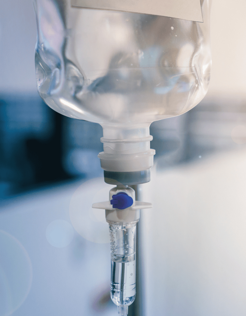


Fluid therapy in clinical medicine is used to fulfill the following objectives: (1) replace dehydration deficits (2) maintain normal hydration, (3) replace essential electrolytes and nutrients, and (4) serve as a vehicle for infusing of certain intravenous medications. The methods used to provide fluids often influence the eventual outcome of the case. Therefore, the clinician and staff should familiarize themselves with the pathophysiology of the diseases they treat and how these conditions relate to the various fluids available for general use.
BODY WATER DISTRIBUTION
B-type natriuretic peptide (BNP) is released from the ventricles in response to stretch or stress. With cardiac disease, the proportion of BNP released from the ventricles increases and the amount released is proportional to disease severity. It is released as a prohormone that is cleaved to an active component C BNP and an inactive component NT-proBNP. The active C-BNP mediates vasodilation and diuresis but has a very short half-life, making it a poor target for assay detection. The inactive portion NT-proBNP has a longer half-life and is the target for current diagnostic testing.
Studies evaluating NT-proBNP have generated values that are useful in specific clinical situations, and it is important to interpret any given NT-proBNP concentration in light of the signalment and clinical question to be answered. Based on studies in small breed dogs with heart murmurs, an NT-proBNP concentration of >900 pmol/L (Idexx) is suggestive of heart disease and echocardiography is recommended. In dogs with established mitral valve disease, a value >1500 pmol/L (Idexx) is suggestive of impending congestive heart failure in the next six to 12 months and warrants close monitoring of the patient.
Various studies in Dobermans have suggested a lower cutoff value for the detection and prediction of DCM in Dobermans (Idexx cutoff is >735 pmol/L). NT-proBNP is most accurate for this purpose when combined with Holter monitoring. NT-proBNP has also been evaluated for its ability to differentiate cardiac from respiratory causes of dyspnea. A value <900 pmol/L in a dyspneic dog suggests CHF is unlikely while a value >1800 pmol/L suggests CHF is likely.
Furthermore, a value < 270 pmol/L in a dyspneic cat suggests CHF is unlikely, while a value >270 pmol/L is supportive of CHF as the cause of dyspnea. NT-proBNP has also been evaluated in asymptomatic cats at risk for heart disease (e.g. murmur present) and various studies support a cutoff of 100 pmol/L for the detection of heart disease in this population. A snap NT-proBNP test is available from IDEXX. For this test, a snap negative correlates with a value <100pmol/L, and a snap positive correlates with >270 pmol/L.
Because there is a “grey zone” with this test, it can be viewed as useful for ruling out moderate to severe heart disease (if the snap is negative) and for detecting moderate to severe heart disease (if the snap is positive); however, it may miss cats with mild heart disease (false snap negative). It is critical that the clinician interpret the NT-proBNP results considering the clinical question to be answered as well as other diagnostic testing.
Indiscriminate testing of animals not at risk for heart disease would be expected to result in a higher false positive rate than has been reported in studies that targeted animals at risk for heart disease. In most situations, a positive test result should prompt an echocardiogram and sometimes thoracic radiographs, blood pressure, and bloodwork as part of further evaluation. Future studies will likely be aimed at determining how NT-proBNP can help guide therapy initiation and monitor response to therapy.
ROUTES OF ADMINISTRATION
Cardiac troponin I (cTnI) is an intramyocardial protein that, when detectable in the bloodstream, indicates myocardial cell injury or death. As such, it is a nonspecific finding that can be associated with many causes of myocardial cell injury including myocardial infarction, myocarditis, cardiomyopathy, congenital heart disease, chronic valve disease, and systemic diseases affecting the heart (e.g. hyperthyroidism, GDV, anemia, toxicities, infections, brachycephalic syndrome, trauma, hypoxia, and renal failure).
The degree of cTnI elevation parallels the extent of myocardial injury and so it can be useful for prognostication. After a single insult, troponin is released within hours, peaks around 24 hours and returns to baseline by two weeks through renal clearance. Serial evaluation, therefore, helps to separate acute from ongoing injury.
Several studies have evaluated cardiac troponin I in dogs and cats with various heart diseases. Low-level elevations (up to approximately 2 ng/ml) are common with congenital and acquired heart disease while massive elevations seem to be most consistent with myocardial infarction or myocarditis. Troponin analysis may have prognostic benefits in some conditions. Troponin was found to be elevated in the majority of dogs with GDV and the value was related to both severity of ECG changes and survival.
Another study found elevated troponin in about half of dogs with motor vehicular trauma and ECG abnormalities in about one-third of these dogs. Cardiac troponin I has also been found to be elevated in dogs before and after cardiac pacing, with a possible myocarditis association. In critically ill ICU patients with no detectable heart disease, troponin elevations were predictive of short-term death.

These studies, among others, highlight that cardiac injury occurs with both cardiac and extracardiac disease and that it can be an important contributor to prognosis. We currently consider cardiac troponin I testing for patients with arrhythmias, especially when the patient is not from a breed known to have a genetically based cardiomyopathy (i.e. a myocarditis screen) and when acute ST segment changes are noted on the ECG. We also use troponin evaluation as part of screening tests for dilated cardiomyopathy and hypertrophic cardiomyopathy with other tests including echocardiography.
We encourage other services within our hospital to consider troponin testing in critically ill patients with GDV, motor vehicular trauma, or other systemic diseases with potential for cardiac involvement. Analyzers capable of detecting lower troponin values (high sensitivity cTnI analyzers) are also available and results suggest prognostic information can be gained in dogs with degenerative mitral valve disease and other conditions. This has also been demonstrated in human medicine and will likely be an area of further study. An elevated troponin concentration should prompt the clinician to pursue further diagnostics including echocardiography, ECG or Holter monitoring, blood pressure, bloodwork, thyroid evaluation and sometimes infectious disease testing.
FLUID SELECTION – CRYSTALLOIDS
Parenteral fluid packages provide a list of the solute content and osmolality. This information is essential when deciding which fluid to use in each clinical situation. Fluids can be conveniently classified based on tonicity (see Table 2). Isotonic crystalloid fluids contain the same osmolality as the extracellular fluids (290-310 mOsm/L). They are excellent solutions for rehydration and maintenance needs, mainly because they can be administered intravenously, intraosseously, subcutaneously, and intraperitoneally. The principal effect of crystalloid solutions is to expand the interstitial volume, not the plasma volume.
This is because of the rapid transendothelial equilibration of isotonic solutions between the intravascular and interstitial spaces constituting the extracellular fluid space. Commonly used isotonic solutions include lactated Ringer’s (near isotonic), 0.9% percent (normal or physiologic) saline, Plasmalyte, Ringer’s, and acetated Ringer’s. Intravenous fluids must be viewed as medications that require careful dose adjustment specific to the patient’s needs. Lactated Ringer’s solution (LRS) is a polyionic, isotonic (273 mOsm/L) solution. It is more physiologic than isotonic saline because its electrolyte concentration is similar to that of plasma. The 28 mEq of lactate anions can help reverse metabolic acidosis because they have the potential to convert to bicarbonate ions by the patient’s liver (in the absence of shock).

Although each liter contains 4 mEq of potassium, supplementation of this cation is usually required for the patient’s maintenance needs. More is necessary for treating hypokalemia. Because the bicarbonate derived from the lactate can promote alkalemia, this solution should not be used if the patient has a coexisting metabolic or respiratory alkalosis. Its calcium content can inactivate certain drugs if combined with this fluid (amphotericin, aminocaproic acid, and ampicillin). The calcium content also makes it contraindicated as a diluent for red blood cell transfusions.
LRS is commonly used as a routine rehydrating and maintenance solution, as a plasma space volume expander in treating shock, and as the fluid of choice in acidotic patients. Isotonic crystalloid solutions such as LRS will distribute between the intravascular and interstitial spaces at a 1:3 ratio. So, if 2 liters of LRS are given IV, 500 milliliters will remain in the intravascular space after two hours, while 1,500 milliliters will end up in the interstitial space. Therefore, it is not an optimal fluid for expanding the intravascular space. Because it is iso-osmolar, no osmotic gradient is created, causing no net movement of water from the intracellular space. Note that adding 20 mEq of KCI to one liter of lactated Ringer’s or isotonic saline will increase the solution osmolality by 40 mOsm/L totaling 312 mOsm/L, which is inconsequential.
An acetated Ringer’s solution (isotonic , 295 mOsm/L) is also available. Its uses and restrictions are similar to those for LRS. Theoretically, sodium acetate is preferable to sodium lactate because a significant portion of the latter anion is converted into liver glycogen. In contrast to LRS, acetate ions are not metabolized by the liver but by muscles and other peripheral tissues. In addition, acetate ions are metabolized differently than the lactate ions and require less oxygen for their metabolism to carbon dioxide and bicarbonate ions when treating shock. Acetated solutions were once not recommended for treating ketoacidosis because acetate might theoretically promote acetoacetate production. However, recent literature refutes that conjecture. Current literature suggests resuscitating septic shock patients with either LRS or acetated Ringer’s.
Ringer’s solution is an isotonic saline solution (309 mOsm/L), with potassium and calcium ion concentrations equal to those normally found in blood and extracellular water. The chloride concentration, however, is supraphysiologic (155 mEq/L). Potassium and calcium ions should be added when patients are depleted of these cations.
“Physiologic,” 0.9% saline (NS) is also isotonic (310 mOsm/L) and is commonly used for rehydration. However, due to its supraphysiologic levels of sodium and chloride ions (154 mEq/L), it has not been the preferred fluid for providing maintenance because large volumes can cause hyperchloremic metabolic acidosis, renal vasoconstriction, delayed micturition, and an increase in the incidence of acute kidney injury (AKI) (NEngJMed, Oct 2015). It has a pH of 5.7. This low pH of 0.9% saline appears related to the polyvinyl chloride bags in which it is packaged since the pH of 0.9% saline in a glass bottle is 7.0.
However, today, there appears to be insufficient data to suggest that 0.9% NaCl is unsafe when used as a maintenance solution (NEngJMed, Oct 2015). This solution is used primarily for plasma volume expansion, hyponatremia correction, and potassium chloride supplementation for treating metabolic alkalosis. Once plasma volume deficits are restored to normal, N.S. should not be used on animals with congestive heart failure or other conditions where sodium restrictions are imposed. Adding 20 mEq of KCl to 1 liter of NaCl 0.9% increases osmolality to 348 mOsm/L which is inconsequential. Saline 0.9% is not recommended for resuscitating septic shock patients. The high chloride content has been associated with poor patient outcomes linked to hyperchloremia, including metabolic acidosis, decreased glomerular blood flow, and peripheral vasoconstriction. (Rochwerg B., “Fluid Resuscitation in Sepsis,” Ann Intern Med 2014; 161:347) On the other hand, some nephrologists currently find no contraindication for using saline under such circumstances.
Plasma-Lyte solution (not listed in Table 2) is the most physiologic of the isotonic solutions (294 mOsm/L) because it has a pH of 7.4, magnesium 3 mEq/L, sodium, and chloride contents of 140 and 98 mEq/L, respectively, and potassium 5 mEq/L. Its acetate, and gluconate contents are 27 and 23 mEq/L respectively, and the osmolality is 295 mOsm/L. It does not contain calcium ions, and it is more commonly used as a maintenance solution.
Dextrose 2.5% in 0.45% saline is isotonic (280 mOsm/L). Once rehydration has been accomplished, and normal electrolyte balance has been restored, it is a valuable maintenance solution when supplemented with potassium chloride. Additionally, it is the fluid of choice for patients whose sodium intake is restricted and those who tend to develop hypernatremia with NS (as seen in some azotemic cats with urethral obstruction). It is important to remember that the dextrose component is rapidly metabolized, leaving a hypotonic fluid in the ECF.

The osmolality of hypotonic solutions is less than that of plasma and extracellular water (<290 mOsm/L). A hypotonic fluid is required if there is a renal concentrating defect with ongoing free water losses or to aid in correcting established hypernatremia. The most used product is 5% dextrose solution (D5W; 253 mOsm/L). It is used (1) in patients with hypernatremia because the dilutional effects will lower the serum sodium level, (2) as a carbohydrate source when another polyionic electrolyte solution is used concomitantly, and (3) as a fluid supplement for patients with sodium intolerance. D5W should not be the sole intravenous fluid for maintenance therapy because of the electrolyte depletion states that can occur, including hyponatremia, hypochloremia, hypokalemia, and hypomagnesemia.
Additionally, this solution should not be administered subcutaneously because extracellular electrolytes tend to diffuse down the concentration gradient into the area of hypodermoclysis (Donnan-Equilibrium Effect). When this happens, the subsequent reduction of plasma solute can theoretically lower the circulating blood volume and cause hypotension. D5W should not be given to correct extracellular volume depletion because two-thirds of the infused volume will enter the intracellular space (where it can cause cellular swelling) within the first hour of infusion, therefore, the expanded plasma space is not maintained. Isotonic saline and LRS, however, remain in the extracellular space for at least two hours. Eight percent of administered D5W stays in the intravascular space, whereas with isotonic saline, at least a quarter of the volume administered remains in the intravascular space. Patients with hypoperfusion can convert glucose into lactate, which can cause lactic acidosis.
The maximum rate dextrose can be infused into humans without producing glycosuria is 0.5 g/kg/hr. About 95% is retained when infused at 0.8 g/kg/hr. (from Drugs Facts and Comparisons 2013. Wolters Kluwer Health, St. Louis, MO.). Dextrose solution equivalencies: D-50-W has 50 gm/100 mL = 0.5 g/mL. Give 1.0 mL/kg (= 0.5 gm/kg) IV slow push. D5W = 5.0 g/100 mL = 0.05 gm/mL. Need 10x the dose of D5W in ml to = that given using D-50-W. D-1.25%=0.125 g/mL. Need 40x (mL) the dose of D-1.25-W in mL to = that given using D-50-W.
Hypertonic solutions have a higher osmolality than plasma and extracellular fluid. A commonly used hyperosmolar solution is dextrose 5% in 0.9% saline (560 mOsm/L). This fluid is not truly hypertonic because the dextrose component is rapidly metabolized after it reaches the bloodstream, therefore, leaving the saline component, which is isotonic at 309 mOsm/L. It can be used as a partial maintenance solution once the patient is fully rehydrated. It is best administered slowly through IV. This solution should not be used in a dehydrated animal because it can potentially promote cellular dehydration and intensify hypovolemia by stimulating diuresis before adequate plasma volume expansion has been achieved. This fluid can be used as an energy source and a sodium supplement in the well-hydrated, hyponatremic patient, such as those with Addison’s disease.
Small-volume hypertonic saline solution (3.0% and 7.2%) provides effective initial resuscitation from hemorrhagic shock. Infusion of 3.0 to 5.0 mL/kg of sodium chloride 7.2% (2,400 mOsm/L) solution to a dog in hemorrhagic shock can rapidly increase systemic blood pressure and cardiac output and produce elevated renal, total splanchnic, and coronary blood flow. The total volume infused should not exceed 4 to 5 mL/kg. The initial dose in cats is 3.0 mL/kg. The beneficial effects of hypertonic saline treatment has also been attributed to its action on the cardiovascular system, including vasodilation, increased in myocardial contractility, and redistribution of fluid from the extravascular to the intravascular compartments, leading to a transient rise in circulating volume.
A solution containing 7.2% NaCl exerts eight times the normal osmotic pressure of the body. The resulting increase in osmotic pressure pulls water from the intracellular space as soon as it is infused into the vascular space. No water is recruited from the interstitial space because the capillary endothelial barrier is freely permeable to small ions such as sodium and chloride. Equilibrium with 7.2% NaCl takes only seconds. For every 1 milliliter of hypertonic saline infused, 8 milliliter of water will transfer from the intracellular to the intravascular space.
Hypertonic saline solution (7.0-7.5%) is also commonly used to treat acute brain swelling at a dose of 3-5 mL/kg IV administered over several minutes.
FLUID VOLUME REPLACEMENT
The animal requires fluids for (1) rehydration, (2) maintenance, (3) replacement of insensible loss volumes, and (4) replacement of ongoing loss volumes. The approximate amount of fluid needed to correct dehydration deficits can be assessed from the history, degree of skin turgor, capillary refill time, and pulse rate and quality. The degree of dehydration ranges from 5% to 12% of the body weight, remembering that 1 liter of water weighs 1 kilogram or 2 pounds. Skin turgor assessment can sometimes be misleading in the obese animal because adipose tissue replaces subcutaneous interstitial water, allowing the skin to maintain its elasticity despite negative water balance.
Additionally, old, cachectic patients that have lost skin resiliency may give a false impression of marked dehydration. To clarify the diagnosis in such questionable situations, clinicians can check for the elevated packed red cell volume and total plasma solids accompanying the hemoconcentration caused by volume depletion. This is valid as long as the patient is neither anemic nor hypoproteinemic, conditions that may give near-normal values despite dehydration. Animals that eat and drink normally without any signs of vomiting and diarrhea should not require IV fluids for water supplementation.
The volume of fluid needed to correct dehydration is calculated from either of the following formulas:
- Volume milliliters of fluid needed = % dehydration x body weight pounds x 500
- Volume milliliters of fluid needed = % dehydration x body weight kilograms x 1000
The maintenance volume (50 to 60 milliliters (about 2.03 oz)/kg/day) is the amount normally required in 24 hours by a well-hydrated patient. It includes the normal urine output approximating 1 to 2 ml/kg/hr plus the insensible fluid loss, of 13 to 20 mL/kg/day. The total 24-hour fluid requirement for the dehydrated animal is the sum of maintenance volume and volume required to correct dehydration plus any estimated ongoing losses.
The initial rate and route of fluid delivery depend on the patient’s status. Mildly to moderately dehydrated small dogs (<10 kilograms) and cats that require short-term fluid treatment can be adequately managed with subcutaneous fluids as long as they are normotensive. Any severely (>10%) dehydrated patient should initially receive fluids intravenously. Because of the accompanying vasocollapse, medications injected subcutaneously may not be adequately absorbed into the systemic circulation. For the mildly to moderately hypovolemic patient, it is recommended that one-fourth to one-half of the estimated dehydration deficit be replaced over the first two to four hours, with the remaining dehydration deficit and maintenance isotonic volumes administered over the subsequent 20 – to 22 – hour period.
The amount of subsequent fluid infusion will depend on the patient’s response to treatment. If the patient is markedly hypovolemic (with “thready” pulses, prolonged capillary refill time, recumbent, mentally depressed, and blood pressure <100 mm Hg), the amount of intravenous fluids administered over the first hour should equal one whole blood volume. This is 70 to 90 mL/kg body weight for the dog and about 35 to 45 mL/kg body weight for the cat. When delivering this much volume over a brief period, it is safer to provide fractional bolus doses of fluids every 15 minutes, at doses of 15 to 20 mL/kg for dogs and 7 to 10 mL/kg for cats. Vital signs in such patients must be monitored every 15 minutes, and fluids should be adjusted accordingly when signs of hypervolemia or interstitial edema occur.
Exceptions to the recommended maintenance doses of fluids occur under the following circumstances:
- Oliguria and anuria. After dehydration deficits are replaced, the patient’s maintenance needs include insensible and ongoing losses plus urine output, which should be quantified. Providing full normal maintenance fluid volumes to oliguric and anuric patients can lead to fatal pulmonary edema or pleural effusion due to iatrogenic intravascular fluid overload.
- Polyuria. Polyuric animals require fluid volumes above normal maintenance needs. Examples include post-obstructive diuresis and recovering AKI patients. Failure to provide these volumes can result in a sustained negative water balance if the patient cannot drink. The maintenance needs for polyuria consist of actual urinary losses plus insensible and ongoing losses. Ensuring adequate treatment is made by weighing the patient daily and assessing the physical and laboratory parameters for hydration. An acute loss of 1 kilogram of body weight suggests a 1 liter of fluid deficit.
- Rapid internal shifts of fluid (third spacing) can occur in pancreatitis, extensive burns, enteritis, and gastrointestinal obstructions. In these conditions, the patient’s fluid needs will exceed the usual maintenance volumes by as much as three times.
The assessment of the animal’s fluid requirements must always be made within the pathophysiologic context of the underlying disorder.
About the Author
Michael Schaer, DVM, DACVIM (SAIM), DACVECC

Dr. Michael Schaer received his D.V.M. degree from the University of Illinois in 1970. He then went to the Animal Medical Center (AMC) in New York City, where he served as an intern and a medicine resident between 1970-73. After the residency, he remained at the AMC as a staff member in the Department of Medicine until 1977. He then joined a private small animal practice in New Jersey until late 1978, when he joined the faculty at the University of Florida, College of Veterinary Medicine where he has remained until now.
He has published several papers and book chapters and lectured nationally and internationally. Dr. Schaer is also the author of five textbooks: Clinical Medicine of the Dog and Cat – 1st-4th editions and Clinical Signs in Small Animal Medicine, 1st and 2nd editions. Dr. Schaer functions mainly as a clinician and teacher at UF. His previous UF professional duties included: Professor and Associate Chairman-Department of Small Animal Clinical Sciences, Associate Chief of Staff-Small Animal Hospital, and Service Chief-Small Animal Medicine. Dr. Schaer served as Interim Chair Department of Comparative, Diagnostic, and Population Medicine from February 2017-September 2018. He is currently Professor Emeritus and Adjunct Professor in Emergency and Critical Care Medicine while still teaching actively in the classroom and the critical care unit. Dr. Schaer is board certified in internal medicine (ACVIM) and emergency and critical care (ACVECC). He is on the board of directors of the North American Society of Toxinology and an associate member of the American College of Physicians.