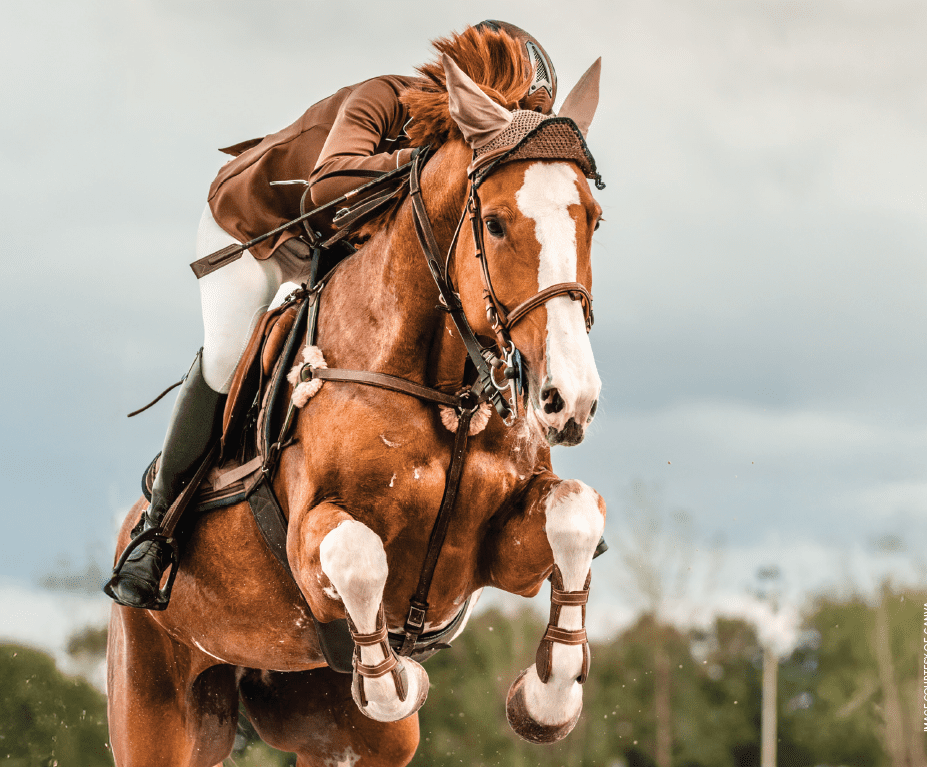
The most common site for lameness issues in sport horses is the distal limb. Many problems are related to chronic repetitive trauma and foot imbalance, while others are more likely of sudden traumatic onset. The source of lameness can often be soft tissues involved in the suspensory apparatus. High-level show jumpers, dressage horses, and Western performance horses often take several years to train to their upper levels, and injuries or illness can substantially affect the time it takes to produce a winning horse. Owners and trainers are concerned that these horses receive the best possible care without excess expense and downtime. The veterinary care of such horses should take an aggressive approach to lameness diagnosis and rehabilitation while the horse is in training, not simply attend to lameness after the horse is no longer able to train. Recognition of early problems involving the suspensory branches may prevent a significant loss of training and competition time as well as extend the horse’s career. Additionally, an accurate diagnosis allows for a more efficient and successful rehabilitation program.
RISK FACTORS
The distal limb plays a significant role in adapting to footing and shock absorption during locomotion. In addition to the corium parietis acting as a ligament within the hoof and the digital cushion functioning in shock absorption, the suspensory apparatus is crucial in absorbing the stress of impact and ground force reaction. Foot balance, both medial to lateral and dorsal to palmar, can have a profound effect on the excursion of joints and the stress on soft tissues of the distal limb. Severely out-of-balance feet (medial to lateral) may therefore result in increased stresses on the suspensory branches. An extremely high heel structure may result in excessive fetlock drop, which in turn stretches the suspensory ligament unit. It has also been demonstrated that an excessively long toe and under-run heel may create unnecessary stress on the suspensory ligament. Significantly toed-in or toed-out structure may abnormally stress one suspensory branch over the other and an over-in-the-knee structure can have some potentially increased suspensory ligament strain. Footing surfaces that are very hard, excessively soft , or unstable can result in aberrant motion and stress that may lead to injury. The demands of high-level sport result in more concussion as well as greater extension and flexion than casual exercise. Fatigue may not be as significant a factor in injuries of the sport horse (with the exception of the eventer) compared to racehorses, but chronic repetitive trauma is thought to be a factor.
Studies of equine degenerative suspensory ligament disease (DSLD) demonstrates that abnormally elevated levels of ligament proteoglycans (PGs) or aggrecans result from changes in the normal homeostatic turnover process. This may also be a factor in otherwise normal ligaments subjected to repeated overload. Repetitive overload may result in disruption of the ligament matrix and promote a low-grade inflammatory reaction as the tissue attempts to repair itself. Studies of horses with degenerative suspensory ligament disease have clearly identified changes in the collagen matrix characteristic of a non-healing wound, with the accumulation of aggrecans and enzymes responsible for their degradation. It has been proposed that ligament failure in DSLD results from a process involving tissue inflammation and the complexation of ADAMTS5, an aggrecanase. It is plausible that a similar process may occur in otherwise normal suspensory ligaments repeatedly subjected to stress and overload. This is yet to be proven in an otherwise normal horse. Tendon and ligament stiffness increases with age and may be a factor in adapting to load changes.
HISTORY AND CLINICAL SIGNS
Horses most often present with unilateral lameness of varying degrees (0-4/5), especially if circled or ridden, which will be discussed later. Subtle lameness requires special efforts to make it more apparent for an objective diagnosis. Many horses vary in their degree of lameness, improving profoundly with a few days’ rest. Mild lameness issues may be best re-examined after a day or two of exercise. Suspensory branch lameness may present in a peracute fashion with significant lameness, local edema, and pain to palpation. However, often signs may be subtle, with the horse having a mild to moderate lameness that changes somewhat with work. Some horses will demonstrate mild thickening in the suspensory branches without apparent lameness, and ultrasound findings may be minimal in early stages of desmopathy. Forelimb lameness may initially present with a mild shortness of stride that “warms out” with ongoing exercise.
Forelimb lameness may be more evident when working in a circle with the affected limb on the outside. Hindlimb issues may mimic more commonly thought-of conditions, such as distal tarsitis or proximal suspensory desmopathy, in that the horse is “weak” behind and “warms out” of the lameness. Some heat or swelling may be perceived in the suspensory branch region, but often this is not the case. Deep palpation of the suspensory branches and forced flexion of the fetlock often produces a painful response. Distal limb flexion most often enhances or produces lameness. Frequently, increased effusion is apparent in the fetlock joint, and the examiner may be tempted to assume it is fetlock synovitis or OA of primary origin. It should be kept in mind that the distal suspensory branch makes up part of the margin of the fetlock joint, and intrasynovial injury may cause synovitis. The current competitive environment in North America often allows for NSAID use in competing horses; medicated horses with mild suspensory branch pain can compete comfortably. However, when Tuesday morning rolls around and the effects of the NSAIDs are gone, the horse may suddenly be more uncomfortable than it was before the event, often with filling and easily identifiable morphological changes. This presents a good opportunity to investigate the case.
PHYSICAL EXAMINATION
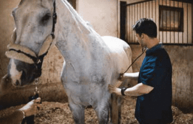
This author believes periodic veterinary inspections of the training sport horse are essential to identify subtle lameness and performance problems before they become serious clinical issues. Although many trainers and owners are very adept at catching subtle soundness and health-related issues, many things may be overlooked by the individual who sees the horse every day. Periodic veterinary inspections should include a general health check and a thorough lameness evaluation. The frequency of these exams will be somewhat dependent on the age and activity of the horse, but two to four times yearly should be a minimum for the competitive horse. Horses with successfully managed orthopedic disease may need to be checked more often.
A typical lameness examination should begin with a thorough visual exam of the horse noting body symmetry and muscle structure. Observing the horse in its stall may provide the examiner with clues regarding the horse’s level of comfort. Watching the horse step out of its stall may give big clues about chronic lameness issues, taking note of shortened or quick steps. After the visual inspection, a palpation examination should be performed. The author uses a modification of an “acupuncture” examination that allows for complete palpation of the horse while eliciting responses from potentially painful areas. Careful inspection of the feet for balance and symmetry should be performed. Flexion manipulations can then be performed in a ‘passive’ sense (without asking the horse to walk or jog away), while noting any resistance or painful responses. It may be appropriate to use hoof testers at this point before starting any exercise. Next the horse can be moved in hand at a walk and trot taking note of any obvious lameness or unusual foot flight or limb motion. This is best performed on hard footing if available. It is useful to see the horse walk in circles as well as on a straight line. Likewise, jogging the horse in circles and in a straight line may provide much more insight into to the horse’s level of comfort. If the horse is too fractious to lunge in circles, jogging in hand on circles may provide an acceptable alternative. The author frequently gives a small dose of sedative (detomidine hydrochloride, 1.5 mg total dose) to evaluate lameness if the horse is not well-behaved. Watching the horse move in straight lines and circles on both hard and soft footing can be immensely valuable in detecting lameness.
The next step is to perform flexion tests of all limbs, with the horse then walking or trotting away. These tests may give clues to soreness that is not otherwise evident. Keep in mind that a positive distal limb flexion may indicate a problem in any number of places, from the distal interphalangeal joint (DIPJ) to even a proximal suspensory ligament issue. It’s a tool for regionalization and augmentation of observation purposes. Care should be taken to not overexert the very lame horse (>3/5) until the nature of the problem is better understood. A detailed record of observations should be maintained for each examination. Following flexion tests, provided the level of lameness is mild, it is advisable to observe the horse work on a lunge line and under saddle. Some horses are difficult to lunge and may pose a hazard for injury to the horse and handler. These horses should be lightly sedated or simply watched under tack. Many lameness conditions are not otherwise apparent until a rider is aboard, or the horse is asked to do more work. The riding examination may give the veterinarian a great deal of information not otherwise apparent during the in-hand exam. Rider weight may change the horse’s balance in a way that augments lameness. Weight on the back may be the source of discomfort and may be demonstrated by a change in the shape of the back, height of head carriage, shortening of stride, or disobedience. The horse should be asked to perform various gaits while under saddle and carefully observed for changes in the level of lameness with each gait, during transitions from one gait to another and at directional changes. Distal limb lameness is often exacerbated by circling, especially on hard footing. Directional changes may enhance this effect, most significantly at the initiation of the change in direction. Suspensory branch issues of a mild nature may be much more obvious under-saddle. Obvious lameness during the undersaddle examination may enable the examiner to perform diagnostic anesthesia more objectively.
At this stage of the examination, the veterinarian should have a good sense of the character of the lameness and may proceed with further diagnostics, such as nerve blocks. Diagnostic nerve blocks are the author’s next diagnostic procedure of choice following the physical examination if they are deemed safe relative to the severity of the lameness. If the degree of lameness suggests further damage may be incurred by exercise, or exercise with desensitized regions, the author will go directly to radiography or, in select cases, ultrasound. Many lameness conditions look alike and flex alike; however, they are not of the same origin. If the lameness is of sufficient grade to produce a clear contrast and has not been reduced by exercise (i.e., warming up), the examiner would be wise to pursue the lameness with diagnostic nerve blocks. Vague lameness issues may be further evaluated by an inertial sensor device, which may give more objective data regarding the response to nerve blocks.
In some cases, physical examination and diagnostic imaging may be sufficient to clearly defi ne the diagnosis such as in the case of significant localized pain and swelling, where nerve blocks may not be required, allowing the veterinarian to proceed with appropriate therapy. Most often, suspensory branch issues will block to a low four-point palmar/plantar nerve block; however, occasionally some will respond to abaxial sesamoid blocks and intra-synovial fetlock anesthesia. Subtle suspensory ligament branch issues may require a combination of diagnostic anesthesia, radiography, ultrasound, and perhaps MRI to fully assess the extent of the injury to provide a better prognosis.
IMAGING
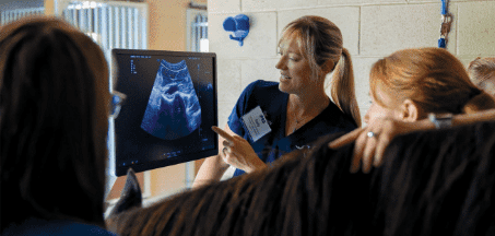
Radiography may be very helpful in the assessment of the fetlock region and suspensory branch issues. Lesions of the proximal sesamoids (so-called “sesamoiditis”) related to the areas of insertion of the suspensory branches may give the examiner a significant clue for the next step in the diagnostic plan. Lesions such as lysis and fragmentation may indicate significant enthesopathies of the suspensory branches. Ultrasound examination is the next step in evaluating problems of the fetlock and distal metacarpal region. A good knowledge of the anatomy is required to properly evaluate the structures, and the suspensory ligament branches can be readily imaged in detail. Lesions occur in various locations along the course of the suspensory branch, but injuries in the middle to distal one-third of the branch are more common in this author’s experience. Lesions may appear as general diff use enlargement and hypoechoic character with no discrete “core lesion,” or as focal anechoic marginal or centralized core lesions.
Many lesions have a linear character and extend along the course of the branch, frequently extending from the surface of the sesamoid proximally. Surface irregularities of the proximal sesamoids are common, reflecting osteolytic lesions of the enthesis. Periligamentous fibrosis and dystrophic mineralization are often seen in more chronic lesions.

Occasionally, nuclear scintigraphy can be useful in further regionalizing the source of the lameness. Areas of increased radiopharmaceutical capture are characteristic of some structures in working horses, and this imaging modality can be extremely helpful in evaluating the significance of the findings from other modalities such as radiography and ultrasound. A suspicious-looking proximal sesamoid on radiography that demonstrates dramatic capture on scintigraphy is thought to have more clinical significance and may reflect a significant suspensory branch enthesopathy.
In recent years MRI has become more accessible to many equine practitioners for imaging lameness cases. While high-fi eld magnets may produce a more detailed image, they currently require general anesthesia for imaging. The standing units are low-field magnets, and image resolution is not as high, but they are nonetheless capable of providing diagnostic images in many, if not most cases of lower limb issues. A specific region should be identified prior to the MRI, as the ability to image multiple regions is limited. MRI provides increased detail of the anatomy and pathology of the fetlock and distal metacarpal region compared to other modalities currently used.
Soft tissue issues not otherwise apparent may be readily visible with MRI. Changes in specific sequence signals related to processes such as bone inflammation are often evident with MRI before any appreciable radiographic changes are visible. The advantage of this is that detecting bone or ligament trauma and inflammation at an earlier stage may allow for more specific treatment modalities and exercise management. The previously discussed imaging modalities are those most often used by this author. Choosing appropriately from this combination of modalities should provide the practitioner specific information that enhances the significance of physical exam findings and local anesthetic blocks, leading to a reasonable diagnosis.

TREATMENT AND REHABILITATION TECHNIQUES
Therapeutic management and rehabilitation for suspensory ligament branch injuries should start with an assessment of the horse’s foot and shoeing status. Shoeing techniques can reduce suspensory ligament loading during initial weight bearing. A wider web at the toe and a narrowed bearing surface in the heel region allows the heel to settle a bit more and the toe to “float,” allowing the DDFT to accept relatively more load. A somewhat wider web of the shoe on the side of the affected suspensory branch is appropriate, unless both branches are significantly affected.

Intra-lesional and perilesional injections of PRP, stem cell preparations, or extracellular matrix preparations (i.e: Regenaflex, Renovo) may be very helpful in encouraging healing with good organization. Th ese products help “direct” healing and recruit progenitor cells. Immediate use of extracorporeal shockwave therapy (ESWT) following intralesional therapy with PRP can increase platelet growth factor release and may further benefit healing responses. More recently, the intralesional and periligamentous injection of alpha-2 macroglobulin (A2M) has been found to reduce chronic inflammation and periligamentous fibrosis in slow-to-respond suspensory branches presumably by inhibition of enzymes.
Application of Class IV laser can encourage the resolution of edema and generation of fibroplasia. Healing is generally slow and requires a gradual rehabilitation program over a minimum of four to six months. Additional stimulation with therapeutic ultrasound every other day for three to four weeks can be of benefit in minimizing edema and stimulating circulatory flow in the affected tissue and various other physical modalities.
RETURN TO ACTIVITY
There is no “cookbook” recipe for rehabilitation of suspensory branch injuries sport horses. Controlled studies of injuries coupled with rehabilitative plans simply have not been done to allow for blanket recommendations. A significant commitment from the owner, trainer and rider is necessary for successful outcomes. Regardless of the nature of the injury, time plays a big part in the rehabilitative process. It must be understood that, while truly unfortunate, some horses will not return to their previous level of performance.
The nature of the tissue injury will dictate much of the rehabilitative process. Early passive mobilization of joints may minimize capsular fibrosis and joint stiff ness, and limited activity may improve comfort in weight-bearing. Significant injuries involving the suspensory branches will require one to three months before any significant activity can begin, and it can be a challenge to regulate the horse’s activity during this phase. Some low-level activity following suspensory injury may be beneficial for healing and may help manage the behavior of the horse. Walking exercise in a water treadmill aft er the first
two months for more serious injuries has been beneficial in this author’s experience. Th is can then be followed by a slow return to walk/trot exercise under saddle, gradually increasing to 10 minutes of trot work over eight to ten weeks. Once trotting ten minutes, canter work generally may be resumed. During this exercise phase, periodic use of ESWT and/or Class IV laser can augment the quality of healing.
Ancillary therapies to help maintain overall condition during “lay-up” must not be overlooked. Daily use of carrot stretch exercises in multiple planes may prove useful for maintaining flexibility of the neck and muscle tone in the back and abdomen. Application of functional electrostimulation (FES) may reduce loss of topline muscle and reduce stiff ness, and there is evidence of the value of FES in horses for increasing mitochondrial concentration in the muscle cells. Mobilization and stretching exercises of the limbs may help minimize joint stiff ness as previously mentioned. Use of vibration plate therapy may also be useful for maintaining back muscle tone. Daily activity and simply moving around and not being “stall bound” have significant effects on the horse’s physiology and well-being.
REFERENCES:
1) Denoix JM, In: Course notes: Module 1, Series 2 The foot and pastern, The International Society of Locomotor Pathology(2008), Middleburg, Va
2) Denoix J.-M., Houliez D. (1997) Déformations du pied à l’appui. In Proceedings ‘Congrès de Podologie Vétérinaires-Maréchaux- Ferrants’. Cluses (France), pp 54-61
3) Nomina Anatomica Veterinaria (Fifth edition) (2012) International Committee on Veterinary Gross Anatomical Nomenclature. Integumentum commune, p. 156-158
4) Lawson SEM, et al, Effect of toe and heel elevation on calculated tendon strains in the horse and the influence of the proximal interphalangeal joint, J. Anat (2007) 210,pp. 583-591
5) Stover, S, Milne Lecture, Proceedings of the AAEP Convention, November, 2022, San Antonio, TX, USA
6) Plaas, A et al, Biochemical identification and immunolocalizaton of aggrecan, ADAMTS5 and inter-alpha-trypsin–inhibitor in equine degenerative suspensory ligament desmitis, Journal of Orthopaedic Research, Vol 29, Issue 6, June 2016, pp. 900-906
7) Dyson S, Greve L, Subjective gait assessment of 57 sports horses in normal work: a comparison of the response to flexion tests, movement in hand, on the lunge and ridden; JEVS 38 (2016) pp 1-7
8) Mitchell, RD, The Role of Ridden Lameness Evaluation, Proceedings of BEVA Convention, Sept 2015, Liverpool, UK
9) Mitchell RD, Distal limb lameness in the sport horse-a clinical approach to diagnosis, Proceedings Ocala Equine Conference, January 2016
10) Dyson SJ, The swollen limb. In: In: Diagnosis and Management of Lameness in the Horse, Eds, Ross M and Dyson SJ, Saunders, Philadelphia, pp 150-151
11) Dyson SJ, Martinelli MJ, (2003) Image description and interpretation in musculoskeletal scintigraphy. In: Equine Scintigraphy, Eds: SJ Dyson, RC Pilsworth, AR Twardock, M Martinelli, EVJ, Newmarket, Suffolk, pp.89-91
12) Greet T, Foreword. In: Equine MRI,(2011) Ed. Murray RC, Wiley- Blackwell, West Sussex, UK, pp. xi-xi
13) Seabaugh, KA, Thoresen, M, Giguere. Extracorporeal shockwave therapy increases growth factor release from platelet-rich plasma in vitro, Frontiers in Veterinary Science, Dec 2017, Vol. 4, article 205
14) Pluim, M et al, High-power laser therapy improves healing of the equine suspensory branch in a standardized lesion model, Front.Vet Sci. 2020 Sep 3; 7:600
15) Rehan AA, Ashan H, and Kahn F, Alpha 2 Macroglobulin: A physiological guardian. J. Cell Physiol. 2912 Aug 228 (8): 1665-1675
16) Peters, D, Tendon and ligament injury with an eye on rehabilitation. In: Proceedings. FAEP, October 2014, Hilton Head Island, SC
About the Author
R.D. Mitchell, DVM, DACVSMR, Certified ISELP Member

Richard D. Mitchell, DVM, DACVSMR is a 1974 graduate of the Oklahoma State University College of Veterinary Medicine and is a senior associate at Fairfield Equine Associates in Newtown, CT and Wellington, FL. He has been internationally certified in veterinary acupuncture (IVAS) and equine locomotor pathology (ISELP), he is a certified Radiation Safety Officer, and he completed requirements for Diplomate status in the American College of Veterinary Sports Medicine and Rehabilitation (ACVSMR) in 2015. Rick served as an official veterinarian to the US Equestrian Team at six Olympic Games between 1992 and 2016. He has a particular interest in lameness diagnostics and imaging. Rick was recognized as a distinguished life member of the American Association of Equine Practitioners and as a distinguished alumnus of Oklahoma State College of Veterinary Medicine in 2022.